Just a few months after German physicist Wilhelm Conrad Roentgen discovered X-rays in 1895, researchers were putting the technology to use to map the heart and its interior anatomy in living subjects.2
And when Enrico Salvioni invented the first iteration of the fluoroscope (which used X-ray techniques to visualize inside the body) a year later, many hailed it as a miracle breakthrough in modern medical technology. However, after the first deaths and injuries from radiation exposure, the X-ray craze died down. In 1904, clinicians adopted protections such as lead shields, aprons and gloves, which are used to this day to help minimize risk.
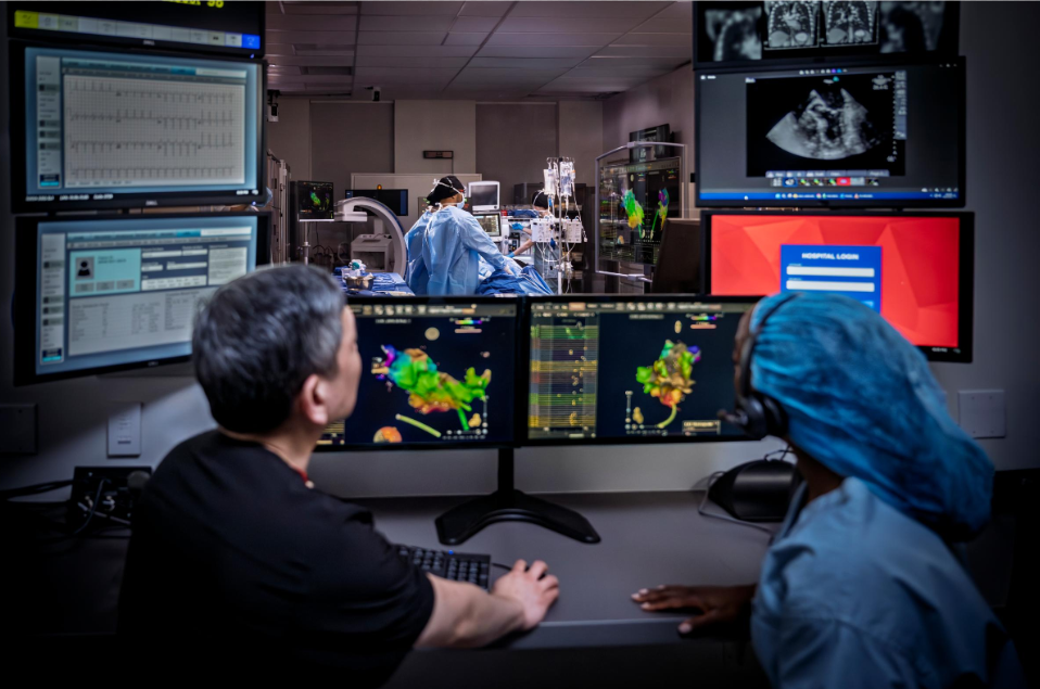
Today's fluoroscopy is different than this early iteration: It takes continuous X-ray images to allow clinicians to view the movement of an instrument, body part or contrast dye inside the body—more like a video. Fluoroscopy is especially useful and important in cardiac catheterization laboratories (cath labs), where it allows for real-time imaging of the heart during procedures. The success of highly complex, minimally invasive electrophysiology (EP) procedures like catheter ablation and left atrial appendage closure depend on high-quality visualization tools that enhance safety and effectiveness.
“Fluoroscopy allows you to visualize what you’re doing in the heart in real time with a full view of the organ,” says Tushar Sharma, Senior Director and Head of Medical Affairs and Preclinical Research at Johnson & Johnson MedTech. “It’s not just a single snapshot, which wouldn’t be enough for sophisticated procedures that we do in the cath lab. You need continuous monitoring because you're treating in a minimally invasive way with no direct visualization of what you’re doing inside the heart.”
Fluoroscopy exposure varies—and can be minimized
While fluoroscopy plays a crucial role in visualizing the heart during complex EP procedures, it also puts physicians and lab staff at risk, especially with longer procedures that create more radiation exposure.3 While patients are also exposed to radiation, healthcare professionals working in cath labs face more repeated exposure that can potentially be hazardous.4 For example, during fluoroscopy procedures, the dose rate of X-rays at the operator's position can range from 1 to 10 mGy/hr. Long-term exposure to this type of radiation can lead to serious health consequences.5
“Clinicians are spending anywhere between 8-12 hours in a cath lab on a daily basis, and if you start doing 15-30 minutes of radiation per procedure, with five procedures a day, that’s close to an hour of radiation exposure a day,” Sharma says.
Official safety legislation was passed in England and the United States in 1921. Now, regulatory and health organizations classify hazards of fluoroscopy as either indirect or direct. A few examples of the latter: an increased risk of cancer, cataracts and reproductive issues in both men and women.
Indirect hazards include problems arising from protection measures. When doing fluoroscopy, clinicians must wear heavy lead jackets for radiation protection.
“Clinicians perform these procedures while standing, which can cause significant back problems,” Sharma says. “So much so that people have had to cut their careers short because of the orthopedic problems caused by these jackets.”
To mitigate these risks, some hospitals have started using lighter lead jackets or zero-gravity systems, which are lead curtains that the physician stands behind to avoid the lead jacket, he explains. And while these techniques minimize exposure, “they don’t eliminate the need for radiation or use of fluoroscopy.”
Advances that are lowering—and will perhaps someday eliminate—fluoroscopy in the cath lab
Johnson & Johnson MedTech’s integrated product portfolio has advanced minimally invasive procedures by combining several key technological advances. Real-time treatment feedback enhances safety while reducing radiation dependence, improving safety for both patients and physicians.
“The main driver for bringing fluoroscopy times down has been the advent of 3D electroanatomical mapping systems,” Sharma says. “In fact, a majority of catheter ablation procedures are already using around 5-7 minutes of fluoroscopy—which is low—thanks to these mapping systems, which integrate AI6 and machine learning capabilities7,8 to help improve efficiency and safety.”
Not to mention, notes Sharma, “the mapping system not only minimizes the need for fluoroscopy, but it also provides better visualization.”9 When doctors manipulate catheters inside the left or right side of the heart, they must know exactly where they are to avoid damaging living structures, he continues. Interpretation can get difficult with fluoroscopy because it only provides a 2D image.
“But a 3D mapping system gives a 3D view of the anatomy, which really allows you to better understand and get oriented to where you are inside the heart,” he says.
With advanced mapping and ablation catheters integrated with Johnson & Johnson’s 3D heart mapping system, doctors can target cardiac tissues that cause irregular heartbeat (such as Afib) and deliver safei and efficient i procedures.
The system incorporates enhanced signal analysis, improved substrate characterization and advanced catheter location to deliver better results while reducing fluoroscopy time.1,8
One technology allows clinicians to take an ultrasound image from the chamber of the heart in a few rotations, and using Deep Learning, creates an image of the shell of the organ without moving the device around to manually contour the heart.
Other imaging technologies, such as ultrasound, have also helped minimize the use of fluoroscopy, “so that in some cases fluoroscopy is often being used as a sanity check rather than as a primary source of visualization,”10 Sharma says. Intracardiac ultrasound, also called intracardiac echocardiography (ICE), for instance, involves inserting catheters into the heart to produce high-resolution images for advanced visualization.11
Johnson & Johnson MedTech makes intracardiac ultrasound catheters that fully integrate with its 3D mapping system. One is enabled with four-dimensional intracardiac echocardiography (4D ICE) sensors that allow it to be visualized on the mapping systems. Another harnesses a new ultrasound crystal that gives very high-quality 2D and 3D images.12,13
Another technology allows clinicians to take an ultrasound image from the chamber of the heart in a few rotations, and using Deep Learning, creates an image of the shell of the organ without moving the device around to manually contour the heart, Sharma says.ii
“This significantly saves time—5-10 minutes of a procedure—since it removes the need to drive the catheter to all nooks and crannies of the heart chamber.”iii
It also gives additional information, like how hard you’re pushing on the heart wall with a catheter.14* “It can tell a physician whether they’re delivering treatment or not,” Sharma says. “Using sophisticated modules that analyze the electrical signals, it can help the physician understand whether they made an impact or not.”
All these innovations benefit clinicians as well as patients: saving timeiv can help doctors deliver more care to as many patients as possible.
“Now the physician has information about what the electrical activity looks like and where they should ablate.15 The combination and integration of all these capabilities,12,13 visualization7 and real-time feedback give us an edge in the electrophysiology space,” Sharma says.
References:
1UG-5407-018H – ELEVATE Module Instructions for Use and Release Notes
2Abrams HL. History of cardiac radiology. AJR Am J Roentgenol. 1996 Aug;167(2):431-8. doi:
10.2214/ajr.167.2.8686621. PMID: 8686621.
3Kosiuk J, Fiedler L, Ernst S, Duncker D, Pavlović N, Guarguagli S, Stegmann C, Miskowiec D, Garcia R, Russo V, Yakushev A, Szegedi N, De Potter T. Fluoroscopy usage in contemporary interventional electrophysiology: Insights from a European registry. Clin Cardiol. 2021 Jan;44(1):36-42. doi:
10.1002/clc.23411. Epub 2020 Nov 21. PMID: 33220000; PMCID: PMC7803367.
4Rizik DG, Rizik MB. Zero Radiation in the Cardiac Catheterization Laboratory: An Aspirational Goal or Moral Imperative? J Soc Cardiovasc Angiogr Interv. 2023 Oct 19;3(1):101131. doi:
10.1016/j.jscai.2023.101131. PMID: 39131981; PMCID: PMC11307473.
5Park S, Kim M, Kim JH. Radiation safety for pain physicians: principles and recommendations. Korean J Pain. 2022 Apr 1;35(2):129-139. doi: 10.3344/kjp.2022.35.2.129. PMID: 35354676; PMCID:
PMC8977205.
6CARTOSOUND™ FAM Module Instructions for Use. UG_5462-018H. October 2023 7 UG-5407-018H - ELEVATE Module Instructions for Use and Release Notes. 8 ‡8 UG-5400-008H - Instructions for Use Software Version 8.1
7UG-5407-018H - ELEVATE Module Instructions for Use and Release Notes.
8UG-5400-008H - Instructions for Use Software Version 8.1
9 UG-5407-018H - ELEVATE Module Instructions for Use and Release Notes
10 Shalom NE, Gong GX, Auster M. Fluoroscopy: An essential diagnostic modality in the age of high-resolution cross-sectional imaging. World J Radiol. 2020 Oct 28;12
11Tong Hu, Tongshuai Chen, Kellina Maduray, Wenqiang Han, Jingquan Zhong. Intracardiac
Echocardiography: An Invaluable Tool in Electrophysiological Interventions for Atrial Fibrillation and Supraventricular Tachycardia. Rev. Cardiovasc. Med. 2024, 25(6), 191. https://doi.org/10.31083/j.rcm2506191
12 Akerström F, Drca N, Urstad MJ, Braunschweig F. Feasibility of a novel algorithm for automated reconstruction of the left atrial anatomy based on intracardiac echocardiography. Pacing Clin Electrophysiol. 2022 Nov;45(11):1288-1294.
13 Di Biase L, Zou F, Lin AN, et al. Feasibility of Three-Dimensional Artificial Intelligence Algorithm Integration with Intracardiac Echocardiography for Left Atrial Imaging During Atrial Fibrillation Catheter Ablation. Europace. 2023 Aug 2;25(9):euad211.
14UG-5407-018H - ELEVATE Module Instructions for Use and Release Notes
15 UG-5407-018H - ELEVATE Module Instructions for Use and Release Notes
ii When comparing SOUNDSTAR™ CRYSTAL Ultrasound Catheter with ACUNAV™ Ultrasound Catheter or SOUNDSTAR™ Catheter
iii Based on Siemens' internal report CRF data responses as of 8/9/2024. CRF Data PN 11291187-EPT-021.
iv Based on Siemens' internal report CRF data responses as of 8/9/2024. CRF Data PN 11291187-EPT-021.
Always verify catheter tip location using common clinical practice for real-time verification (inspection of IC signals, direct imaging guidance such as fluoroscopy or ultrasound, etc.) and consult the CARTO™ 3 System User Guide regarding recommendations for fluoroscopy use.
Canpolat, U. et al (2020). State of Fluoroless Procedures in Cardiac Electrophysiology Practice. J Innov Cardiac Rhythm Management. 11(3), 4018–4029. Sommer, P. et al (2018) Safety profile of near-zero fluoroscopy atrial fibrillation ablation with non-fluoroscopic catheter visualization: experience from 1000 consecutive procedures, EP Europace, Volume 20, Issue 12, Pages 1952–1958.
Important information: Prior to use, refer to the instructions for use supplied with this device for indications, contraindications, side effects, warnings and precautions.
Caution: US law restricts this device to sale by or on the order of a physician.
US_ELP_SOEP_406674
© Johnson & Johnson and its affiliates 2025