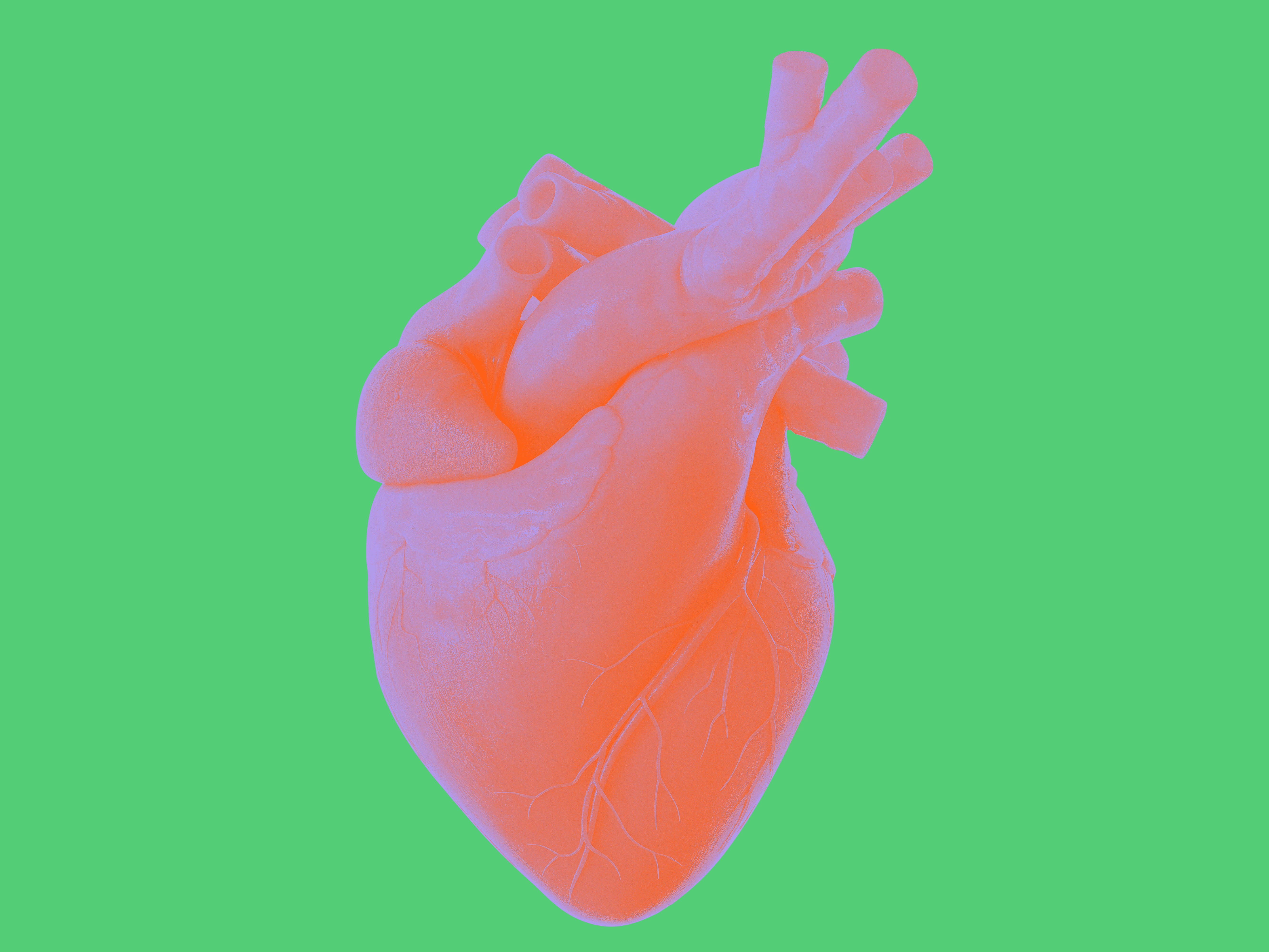
What’s in an intravenous catheter? Micro miracles of embedded technologies that provide electrophysiologists with clarity when repairing the heart during minimally invasive surgery.
Technology is increasingly enabling precision repairs to the human body without disturbance to tissue or bone structure and can deliver greater efficacy than medications for conditions such as atrial fibrillation — the most suffered cardiac arrhythmia. A recent study showed that this debilitating condition affects more than 37 million people throughout the world, and its prevalence in the US alone is predicted to be as high as 12 million people by 20501.
Electrophysiology — the medical specialty that primarily treats cardiac arrhythmias — is a leading field in the deployment of medical technologies that enter the body via a small incision. Once inside the body, they provide detailed images of their pathway and surroundings, allowing electrophysiologists (EPs) to confidently navigate and carry out targeted procedures such as catheter ablation.
Catheter ablation is the selective disabling of rogue electrical impulses that cause the heart to beat irregularly, which typically causes symptoms such as dizziness, shortness of breath and anxiety. These cardiac arrhythmias also contribute to mortality by increasing the likelihood of stroke (blood that is not constantly moving can clot and move to the brain) and cardiac arrest.
Very simply, ablation is carried out using a fine catheter — around 2.64 millimeters in diameter — which is fed up through a vein, usually in the groin, to the heart. Inside the heart, its sensors identify the path of disruptive electrical activity. Most commonly, the tip of the catheter is used to deliver heat in the form of radio frequency, which destroys individual cells in the heart muscle so that the electrical impulse can no longer pass through. Stopping the passage of irregular electrical activity allows the heart to return to its natural pace, or sinus rhythm.
Although medication is still the most applied method of moderating cardiac arrhythmias, great strides have been made in effectively stopping the most common arrhythmia — atrial fibrillation
(AFib) — using radiofrequency (RF) ablation. Today, new minimally-thermal technologies are also being developed to deliver potentially even safer procedures.
AFib causes fast and irregular heartbeats, usually more than 400 beats per minute, compared to the normal range of 50-150 bpm. It falls into three categories: Paroxysmal AFib (intermittent episodes that stop within seven days), Persistent AFib (episodes last longer than seven days) and Longstanding Persistent AFib (episodes lasting longer than a year)2.
“Since Paroxysmal AFib (PAF) treatment was discovered about 25 years ago,” says Avi Shalgi, head of R&D for J&J’s Cardiovascular and Specialty Solutions (CSS) group of companies, “innovations have improved the accuracy of treatments, the ability to control and have better contiguity between lesions, reduction in radiation exposure time and shorter treatment times — which adds up to much better outcomes for patients.”

He adds that in the early days most electrophysiologists could ablate to provide patients with around one year free of PAF in 60% of cases. “Today many EP physicians have PAF efficacy close to 90%, along with very safe and short procedures compared to 20 years ago.”
Shalgi orchestrates the work of around 800 researchers: materials and sensor specialists, software engineers and other technicians based in both Israel and the US. He says, “I love to bring the best solutions to treat patients; to give my customers, the physicians, the tools that help them make the most effective and efficient use of their knowledge and skills.”
Here he lists five technologies that have “transformed the field of electrophysiology of the heart in recent years.” And he gives some insights into how these technologies might soon be applied to other areas of surgical intervention.
1. Intrabody Electromagnetic (EM) navigation
How it works in the heart: Operating inside the body, beyond the line of sight, this technology acts like an internal GPS. “We developed a location technology that can track the position of our tiny EM sensors with an accuracy of less than a millimeter and one degree in 3D space,” says Shalgi.
When embedded in a mapping/diagnostic catheter, EM sensors show exactly where the catheter is inside the body. When combined with other in-catheter technologies, the sensor “acts like a converging system”, that puts other information gathered from within the heart “on one frame of reference”, explains Shalgi, “so you know where each one of them is oriented in space.”
Where to next? Location sensors can be used for navigation in other parts of the body, such as the abdomen, legs, and lungs. Incorporated in other micro tools designed for minimally invasive surgery, electromagnetic navigation means “you don’t need to open the chest or the abdomen to operate — you just need a small incision,” says Shalgi. You can “see” without having a line of sight.
This technology has already been successfully adapted for ear, nose and throat (ENT) procedures facilitated by a Johnson & Johnson MedTech company, Acclarent, which also comes under Shalgi’s R&D purview. For physicians, a virtual view inside the frontal cavities of the skull — using images from CT or MRI scans or a combination of both — allows delicate adjustments to be made, such as shaving of bone to enlarge the sinuses for easier breathing.
2. Real time electrophysiology mapping linked to heart anatomy and structure
How it works in the heart: By integrating and embedding tiny EM sensors in various catheters, technicians can collect internal electrograms from every location in the heart. “So, by moving the catheters around the heart chambers,” says Shalgi, “we generate a full three-dimensional electro anatomical map of the heart showing electrical signals and their precise location.” This enables EPs to understand the arrhythmia behavior — the wave propagation and signals causing the irregular heartbeat — in each patient’s heart, in real time, and treat the arrhythmia.
Where to next? Real-time electrophysiology mapping is unique to signals from the heart, and therefore unlikely to be applied to other parts of the anatomy.
3. Irrigation in the ablation catheter
How it works in the heart: Ablation has enabled the treatment of arrhythmias using radio frequency energy to destroy selected cells in the pathway of rogue electrical pulses — thereby blocking their effect on the heart.
The RF energy is delivered by touching the cells with the tip of the catheter, which heats the tissue and destroys it. “Before we developed an irrigation technique, the tip could become so hot, it would cause charring, coagulation of blood in the vicinity, or even result in a clot,” says Shalgi.
“We have since developed a system of 56 tiny holes in the tip of the catheter — each one is a fraction of a millimeter in diameter — which delivers cooling saline to the site and reduces unnecessary charring and minimizes blood coagulation.”
Where to next? In future, this technology will be useful in any treatment involving ablation by heat sources such as radio frequency.
4. Contact force technology
How it works in the heart: Sensors do a world of heavy lifting in the treatment of AFib via electrophysiology. In this instance, tiny force sensors embedded in the catheter tip assess the quality and consistency of the ablation/lesion being created. Precise mapping capabilities and force sensors, orchestrated by a formula developed at Biosense Webster, Inc. R&D, today enable real-time calculation of the force/time/power used to conduct ablations. “It means EPs can deliver very consistent lesions to the tissue they want to disable, and get much better results than without it,” says Shalgi.
Where to next? “Force sensing is applicable to other ablation procedures and can also be used with a new emerging Pulse Field Ablation technology to deliver very consistent lesions, without excessive heat, where you want to ensure that during ablation, you’re not perforating the tissue,” Shalgi predicts. “Control of force will be important in other surgeries.”
5. Real-time imaging (using real-time ultrasound)
EPs are still using computed tomography (CT) and magnetic resonance imaging (MRI) to generate images of the heart prior to catheter ablation, says Shalgi, but these technologies have some downsides in the treatment of common conditions such as AFib: they slow down a patient’s journey to treatment because the patient needs to come in prior to the procedure in order to have the scan; CT also adds to the potentially dangerous radiation burden for any patient; and such scans are expensive to the health system.
Over the years, in collaboration with companies known for medical imaging, Biosense Webster, Inc. has developed “a disruptive technology”, says Shalgi, embedding intracardiac echocardiography (ICE) navigation in its catheters, which generates real-time ultrasound images of the heart from within.
“Two years ago, we also acquired a new company that had leveraged a 3D transducer element, which shows you sections of the heart in 3D — you see the heart moving,” says Shalgi.
“By using the navigated ICE catheter, and combined ultrasound images,” he continues, “EP physicians can generate a very accurate anatomical map of the heart’s chambers. You don’t need pre-imaging because you can see what you’re doing in real time.”
Real-time in-body imaging reduces the requirement for prior CT scans, and the need for X-rays to guide the ablation procedure, thereby reducing the overall radiation load that both patients and EPs are exposed to and minimizing the patient’s journey to treatment.
Where to next? Ultrasound can be used in other parts of the body, with its only drawback being that it needs a fluid environment. Says Shalgi, “If you have air, for example, as in the lungs, ultrasound doesn’t work, because its waves are reflected in air.” Other intra-body environments that may benefit in the future from catheter-generated real-time imaging, include orthopedic surgeries, particularly the treatment and repair of muscles.
The catheter is a big system in a slender package
An EP procedure can involve several catheters simultaneously deployed in the heart — for example, a diagnostic catheter, an ablation catheter, and an ultrasound-imaging catheter. Coordinating catheters and relaying their feedback, is Biosense Webster, Inc.’s CARTO® 3 System. Its integrated hardware and software collate every detail of the procedure underway in one frame of reference — real-time imagery of the heart. “Our catheters are a full system made miniature,” says Shalgi. CARTO® 3 System orchestration puts electrophysiologists in control of a suite of micro tools: “It's like the full cockpit of an aircraft the pilot sees everything in front of them and can make informed decisions.”
Important information: Prior to use, refer to the instructions for use supplied with this device for indications, contraindications, side effects, warnings and precautions. Caution: US law restricts this device to sale by or on the order of a physician
The THERMOCOOL SMARTTOUCH® SF Catheter is indicated for the treatment of drug refractory recurrent symptomatic paroxysmal atrial fibrillation (AF) and for drug refractory recurrent symptomatic persistent AF (continuous AF > 7 days but < 1 year), refractory or intolerant to at least 1 Class I or III AAD, when used with the CARTO® 3 System.
1 Lippi G, Sanchis-Gomar F, Cervellin G. Global epidemiology of atrial fibrillation: An increasing epidemic and public health challenge. Int J Stroke. 2021;16(2):217-221.
2 What is Afib? Johns Hopkins Medicine website. https://www.hopkinsmedicine.org/health/conditions-and-diseases/atrial-fibrillation Accessed July 14, 2023.
259893-231011
© Bionsense Webster, Inc. 2023